PA OBLIQUE THUMB PROJECTION
45° oblique projection of the first digit for complete three-dimensional evaluation
OBLICUA
Proyección PA Oblicua de Pulgar

PA
Proyección PA de Pulgar para comparación
COMPARISON: PA vs OBLIQUE THUMB
STANDARD PA
- Flat posteroanterior view
- Overlapping of structures
- Limited joint evaluation
- Difficult to spot non-displaced fractures
45° OBLIQUE
- Three-dimensional view
- Better joint space visualization
- Detailed view of the 1st Metacarpal
- Ideal for Bennett/Rolando fractures
Exposure Factors
45-50
Kilovoltage (kV)
2-4
mAs
100 cm
Distance (SID)
Fine
Focal Spot
No
Grid (Bucky)
Patient Positioning
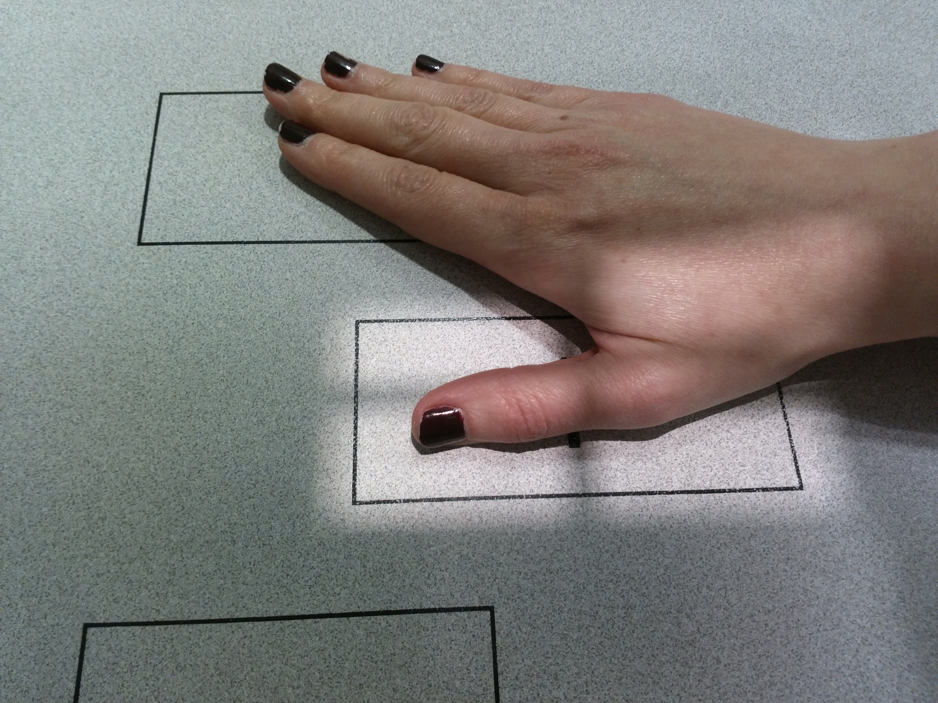
Place hand in prone position (palm down) on the IR.
Rotate the hand slightly until the thumb is in a natural 45° oblique position.
The thumb must be fully extended and parallel to the IR.
Center the thumb on the image receptor area.
Central Ray
1st MCP Joint
Location: Perpendicular to the first metacarpophalangeal (MCP) joint.
Visible Anatomy
Phalanges
Distal and proximal phalanges visible.
1st Metacarpal
Visualized in its entirety.
Joints
IP and MCP joint spaces open.